What are X-Rays?
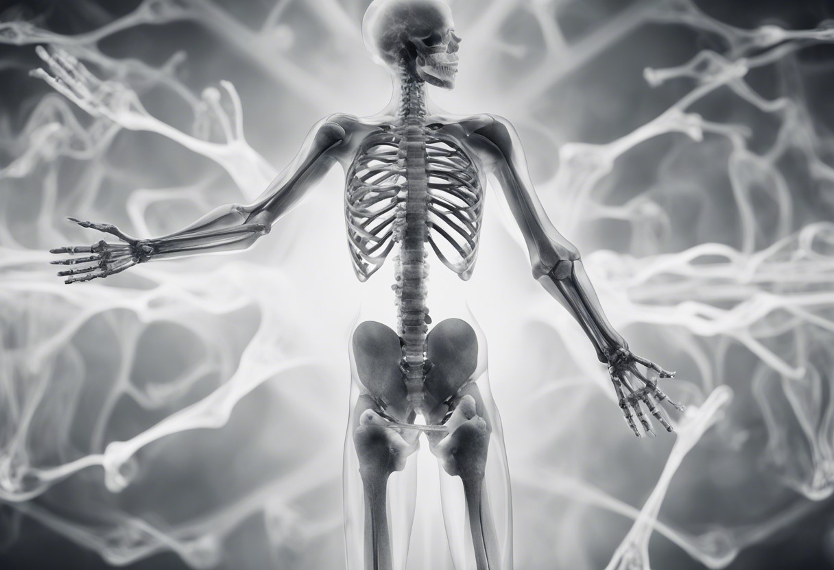
X-rays are a form of electromagnetic radiation with wavelengths shorter than ultraviolet light. They possess the unique ability to pass through various materials, including the human body, which makes them invaluable in medical diagnostics. X-ray imaging is one of the most common and essential tools in modern medicine, enabling healthcare professionals to visualize the internal structures of the body to diagnose and monitor a wide range of conditions.
How Do X-Ray Devices Work?
X-ray devices function by emitting a controlled beam of X-rays from an X-ray tube, which then passes through the body. Different tissues absorb X-rays at different rates; dense materials like bones absorb more X-rays and appear white on the resulting image, while softer tissues allow more X-rays to pass through and appear in shades of gray. The X-rays that pass through the body are captured on a detector or film, creating an image that can be analyzed by medical professionals.
Main Parameters of X-Ray Devices
- X-Ray Tube Voltage (kVp):
- Description: The kilovolt peak (kVp) determines the energy and penetrating power of the X-ray beam. Higher kVp values increase the energy of the X-rays, allowing them to penetrate denser tissues.
- Significance: Higher kVp settings are used for imaging thicker or denser body parts, such as the chest or abdomen, while lower kVp settings are suitable for extremities and soft tissues. Adjusting the kVp is crucial for optimizing image contrast and reducing patient dose.
- Tube Current (mA):
- Description: Measured in milliamperes (mA), the tube current controls the number of X-rays produced. Higher mA settings increase the number of X-ray photons generated, resulting in a more intense beam.
- Significance: The mA setting affects the image’s brightness and noise levels. Higher mA settings produce clearer images but also increase the patient’s exposure to radiation. Balancing mA is essential for achieving high-quality images while minimizing radiation dose.
- Exposure Time (s):
- Description: Exposure time refers to the duration for which the X-ray beam is active, measured in seconds. It determines how long the X-rays interact with the body and the detector.
- Significance: Longer exposure times can enhance image quality by allowing more X-rays to reach the detector, but they also increase radiation dose. Shorter exposure times are preferred to minimize motion artifacts and patient exposure.
- X-Ray Beam Collimation:
- Description: Collimation involves shaping and directing the X-ray beam to the area of interest, reducing the exposure to surrounding tissues. Collimators are adjustable devices that limit the size and shape of the X-ray field.
- Significance: Proper collimation enhances image quality by reducing scatter radiation and improving contrast. It also protects patients by limiting radiation exposure to only the necessary area.
- Detector Type:
- Description: X-ray detectors capture the X-rays that pass through the body and convert them into visible images. Common types include film, computed radiography (CR) plates, and digital radiography (DR) detectors.
- Significance: The choice of detector affects image quality, workflow efficiency, and radiation dose. Digital detectors (DR) provide superior image quality, faster processing times, and lower radiation doses compared to traditional film.
- Image Resolution:
- Description: Image resolution refers to the clarity and detail of the X-ray image, typically measured in line pairs per millimeter (lp/mm). Higher resolution allows for better visualization of small structures.
- Significance: High-resolution images are crucial for accurately diagnosing conditions, particularly in areas requiring detailed views, such as dental imaging or detecting microfractures.
- Radiation Dose:
- Description: The radiation dose is the amount of ionizing radiation absorbed by the patient during an X-ray examination, measured in millisieverts (mSv).
- Significance: Minimizing radiation dose is vital to ensure patient safety while maintaining diagnostic image quality. Techniques such as adjusting kVp, mA, and exposure time, along with using advanced detectors, help achieve this balance.
Types of X-Ray Devices
- Conventional X-Ray Machines:
- Description: These are standard X-ray units used for a variety of diagnostic purposes, including chest, abdominal, and skeletal imaging.
- Applications: General diagnostics, emergency imaging, and routine examinations.
- Fluoroscopy:
- Description: Fluoroscopy provides real-time X-ray imaging, allowing dynamic studies of the body’s internal functions. It’s often used with contrast agents to enhance visibility.
- Applications: Gastrointestinal studies, angiography, and interventional procedures.
- Computed Tomography (CT) Scanners:
- Description: CT scanners use multiple X-ray beams and detectors to create cross-sectional images of the body, providing detailed three-dimensional views.
- Applications: Detailed imaging of internal organs, detecting cancers, and planning surgeries.
- Mammography:
- Description: Specialized X-ray equipment designed for breast imaging, using low-dose X-rays to detect breast cancer at early stages.
- Applications: Breast cancer screening and diagnosis.
- Portable X-Ray Units:
- Description: These compact and mobile X-ray machines are used for bedside imaging, particularly in emergency rooms and intensive care units.
- Applications: Imaging for immobile patients, emergency diagnostics, and field medical applications.
The Future of X-Ray Technology
Advancements in X-ray technology continue to enhance diagnostic capabilities while prioritizing patient safety. Innovations such as digital tomosynthesis, dual-energy X-ray absorptiometry (DEXA), and artificial intelligence (AI) integration are set to revolutionize medical imaging. These developments promise greater accuracy, faster results, and reduced radiation doses, ultimately improving patient care and outcomes.


